Rice lab finds molecular clues to Wilson disease
Using a combination of computer simulations and cutting-edge lab experiments, physical biochemists at Rice University have discovered how a small genetic mutation -- which is known to cause Wilson disease -- subtly changes the structure of a large, complex protein that the body uses to keep copper from building up to toxic levels. "The protein we study is like a big puzzle," said lead author Agustina Rodriguez-Granillo, the Rice doctoral student in biochemistry and cell biology who carried out the mathematical simulations and laboratory research. "The mutation that causes most cases of Wilson disease is well-known, but our study looks at the overall puzzle to see how such a small mutation can alter the shape and function of such a large and complex protein."
The protein in question is called ATP7B, which is a multidomain protein that sits in an internal membrane and regulates the movement of copper atoms inside human cells. Though large quantities of copper can be toxic, our bodies need a small amount for key enzymes involved in, for example, respiration and brain functions. ATP7B acts something like a warehouse manager, locking up bulk quantities of copper and handing it out for use in these proteins.
Wilson disease is a genetic disorder that alters the ATP7B protein's ability to work, causing copper to build up to toxic levels in the liver, brain, eyes and other organs. Over time the disease can cause life-threatening organ damage. Wilson disease affects as many as 150,000 people worldwide.
The new study is available online from the Journal of Molecular Biology. It focused on the genetic flaw that causes most cases of Wilson disease. That flaw, known as H1069Q, is caused when just one out of the more than 1,400 amino acids in ATP7B is changed. That amino acid is a histidine located at position 1069. In the disease-causing form of the protein, this histidine is replaced with a glutamic acid.
"This mutation occurs at a crucial location where the protein typically binds with a molecule called ATP that provides the energy the protein needs to move copper from place to place," said study co-author Pernilla Wittung-Stafshede, an adjunct professor of biochemistry and cell biology at Rice and Rodriguez-Granillo's adviser. Wittung-Stafshede, professor in chemistry at Umea University in Sweden, said, "Past studies have compared the behavior of the mutant protein with that of the nonmutant and found very little difference, so it was unclear how this small change led to the devastating effects that are seen in Wilson disease."
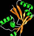
Using a combination of experimental data and computer simulations that looked specifically at a portion of the protein called the N-domain, where the H1069Q mutation occurs, Wittung-Stafshede, Rodriguez-Granillo and postdoctoral researcher Erik Sedlak (now at the University of Texas at San Antonio) confirmed that ATP's function was significantly reduced in the mutant form of the protein. They also found that the mutation caused structural changes in other sections of the protein that were far away from the mutation site. For example, the healthy form of the protein is capped with a large, flexible loop. The purpose of the loop is unknown, but its shape is altered and more compact in the diseased form of the protein.
"This implies that the loop has some importance, perhaps in regulation of ATP7B's activities, and we intend to follow up on this in our future studies," Rodriguez-Granillo said.
Source: Rice University
Other sources
- Molecular Clues To Wilson Disease: How Mutation Alters Key Proteinfrom Science DailyThu, 21 Aug 2008, 21:35:08 UTC
- Rice lab finds molecular clues to Wilson diseasefrom PhysorgTue, 19 Aug 2008, 18:35:14 UTC
