Miniature brains made from patient skin cells reveal insights into autism
Understanding diseases like autism and schizophrenia that affect development of the brain has been challenging due to both the complexity of the diseases and the difficulty of studying developmental processes in human tissues. In a study published July 16 in Cell, researchers have made steps toward overcoming these challenges by converting skin cells from autism patients into stem cells and growing them into tiny brains in a dish, revealing unexpected mechanisms of the disease. Most autism research has taken the approach of combing through patient genomes for mutations that may underlie the disorder and then using animal or cell-based models to study the genes and their possible roles in brain development. Although this has yielded a handful of rare disease genes, the limitations of these models and the complexity of the disorder have frustrated researchers and left over 80% of autism cases with no clear genetic cause. The new study now turns the traditional approach on its head.
"Instead of starting from genetics, we've started with the biology of the disorder itself to try to get a window into the genome," says senior author Flora Vaccarino the Harris Professor of Child Psychiatry and Professor or Neurobiology at the Yale School of Medicine.
The clinical characteristics of autism are complex and wide-ranging, making the prospect of finding common underlying factors slim. To stack the deck in their favor, the researchers focused on the approximately one-fifth of autism patients that share a distinctive feature correlated with disease severity--an enlarged brain. After isolating skin cells from these individuals, as well as their unaffected fathers to provide a point of comparison, the researchers converted the cells into induced pluripotent stem cells that were then grown into miniature brains.
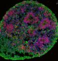
These so-called "brain organoids" are just a few millimeters in diameter but mimic the basics of early human brain development, roughly corresponding to the first few months of gestation. When the researchers analyzed the patient organoids, they uncovered altered expression networks for genes controlling neuronal development. Patient organoids showed an unexpected overproduction of inhibitory neurons that quiet down neural activity, while those that excite the partners they're wired to were unaffected, leading to an imbalance in neuron type. Remarkably, by suppressing the expression of a single gene whose expression was abnormally increased in patient organoids, the authors were able to correct this bias, suggesting that it may be possible to intervene clinically to restore neuronal balance.
With current technology, human brain organoids only recapitulate early stages of development; however, efforts to extend their growth to later stages are under way by a number of groups and will allow even further insights into disease mechanisms. The authors are now using their data to home in on the difficult-to-find mutations or epigenetic changes responsible for the gene expression alterations and neuronal imbalance observed in the study.
According to Vaccarino: "This study speaks to the importance of using human cells and using them in an assay that could bring a better understanding of the pathophysiology of autism and with that, possibly better treatments." The success of the approach also suggests that similar methods might be used to gain important insights into other human developmental diseases that have until now been difficult to crack open.
Source: Cell Press
Articles on the same topic
- Making 'miniature brains' from skin cells to better understand autismThu, 16 Jul 2015, 16:37:24 UTC
- Scientists find mechanism for altered pattern of brain growth in autism spectrum disorderWed, 15 Jul 2015, 19:04:26 UTC
- New evidence linking brain mutation to autism, epilepsy and other neuro disordersWed, 15 Jul 2015, 19:04:20 UTC
- MRI studies point to brain connectivity changes in autism spectrum disordersWed, 15 Jul 2015, 19:04:10 UTC
- Older age at onset of type 1 diabetes associated with lower brain connectivity laterWed, 15 Jul 2015, 19:03:53 UTC
- Brain study reveals insights into genetic basis of autismWed, 15 Jul 2015, 19:03:44 UTC
Other sources
- Miniature brain in a dish reveals an outsized secret about autismfrom LA Times - ScienceFri, 17 Jul 2015, 2:00:22 UTC
- Miniature brains made from patient skin cells reveal insights into autismfrom Science DailyThu, 16 Jul 2015, 18:00:39 UTC
- Mechanism for altered pattern of brain growth in autism spectrum disorder discoveredfrom Science DailyWed, 15 Jul 2015, 20:30:36 UTC
- Older age at onset of type 1 diabetes associated with lower brain connectivity laterfrom Science DailyTue, 14 Jul 2015, 15:30:47 UTC
- MRI studies point to brain connectivity changes in autism spectrum disordersfrom Science DailyTue, 14 Jul 2015, 15:00:57 UTC
- Brain study reveals insights into genetic basis of autismfrom Science DailyMon, 13 Jul 2015, 16:10:10 UTC
